How to Measure: Kirby Bauer Test Instructions
Figuring out how to measure Kirby Bauer test inhibition zones is much less difficult than you might imagine. It’s all a matter of understanding the purpose behind it, and then implementing the simple steps to get the job done.
What Is the Purpose of the Kirby Bauer Test?
Bacteriologists began their important work shortly after the discovery Louis Pasteur made with his microscope in the early 1800s. Once scientists realized they could identify the tiny living organisms that had been coexisting with us since the beginning of humanity, investigations took off.
The field of microbiology was born, and an increasing number of scientists grew interested in studying these minute life forms.
From the beginning of time, three primary organisms have lived on earth —- archaea, bacteria, and yeast.
Archaea and bacteria are closely related in that they are microorganisms without an enclosed nucleus. They are distinct in that archaea can survive in extreme temperatures, and they were likely the first signs of life on earth, capable of thriving in both freezing and ultra-hot temperatures.
Bacteria probably evolved from archaea, a less extreme prokaryote. While archaea reproduce through fission, bacteria often produce spores and can be pathogenic. No archaea are known to be pathogenic.
Finally, scientists believe that yeast likely evolved from bacteria as a means of competing for food. In those early, ancient times, living organisms received their food from the sun and the process of photosynthesis. As plant life evolved, yeast evolved to feed from the sugar in plants.
To do this, it developed its own nucleus, becoming a prokaryote.
Evolution of Early Organisms
Now, hundreds of years later, these three life forms are entirely distinct from each other, and scientists have only been studying them for less than 200 years. We know what we know precisely because of microscopes and the development of the Kirby Bauer Test.
In the early days, after microscopes became more widely available, microbiologists would study archaea, bacteria, and yeast using more rudimentary methods.
They would use gelatin to culture these life forms and then cover them with a glass bell jar.
To understand how bacteria behaved, they would watch it grow over time, interacting with various stimuli, early antibiotics or nutrients. The growth or inhibition of bacteria would give clues as to how effective various additives to the gelatin culture were.
Monitoring Bacteria
Eventually, as the late 1800s drew to a close, bacteriologists began to develop a uniform approach to studying bacteria.
They would culture the bacteria on agar agar inside of a Petri dish that came with a lid, a long way from beef gelatin and a bell jar.
Then, they would count the colony forming units to understand how many CFUs appear to the naked eye on the agar.
Get access For Free to our comprehensive How to Count Colonies on Agar Plates PDF Guide (19 pages) put together by our specialists, Dr. Katja Schulze, who specialized in bioinformatics and image recognition of microscopic images, and Dr. Ulrich M. Tillich, who specialized in laboratory automation!
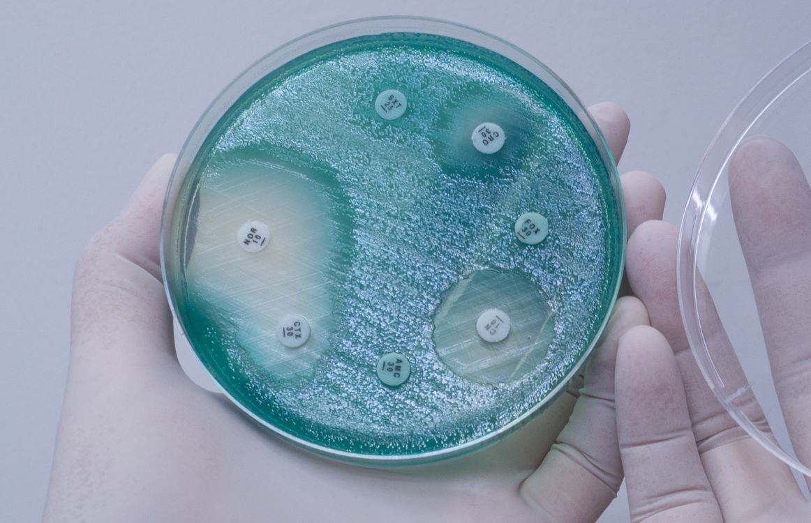
To test antibiotics, a small disk soaked in antibiotics would be placed on the agar, over the bacterial culture, and, if a clear space appeared around the disk, scientists knew the antibiotics were effective. How large this space, or zone of inhibition, was, and how long it lasted, provided information as to just how effective the antibiotic was.
The problem was that, across the globe, different scientists used slightly different techniques. This variance made it difficult for medical and health organizations to communicate and collaborate.
The Development of the Kirby Bauer Technique
In the 1950s, the World Health Organization called for a single, uniform approach to what had previously been known as the disk diffusion test. Bacteriologists around the globe submitted their papers with their specific process, which they thought was best.
Two men, W. Kirby and A. Bauer were the ones who ultimately designed the winning process, so now the disk diffusion test is widely known as the Kirby Bauer Test.
Kirby Bauer Technique
The technique is simple.
First, a bacterial sample is collected and kept in a hygienic vessel, like a vial with an airtight stopper.
Next, the bacterial sample is diluted in the lab to ensure the bacteria does not overwhelm the agar when it is added. Some bacteria can grow so rapidly that it will present (sometimes far) more than 30 colony forming units, which can be difficult, if not impossible, to count (if that also needs to be done).
To dilute the sample, simply extract one part of the sample and add it to a separate vessel, to which you will add 10 parts of saline or filtered water. Then shake gently to ensure the sample is mixed fully with the dilutant.
Then, dilute the already diluted sample at a factor of 1:10 again, to get a 1:100 diluted sample.
Now, with that diluted sample, you will take a sample with a cotton swab and swipe that sample in a tight zigzag pattern across the agar nutrient, from edge to edge of the Petri dish. Turn the plate 90 degrees and repeat this process to ensure the entire agar is covered.
Place the lid on the Petri dish and flip it upside down. Incubate the bacteria according to the instructions of your specific sample.
After the allotted time, remove the sample for a CFU count. At that point, you can place antibiotic disks, ideally using an automated disk dispenser on the agar, over the bacteria. Incubate again according to the bacteria and antibiotic instructions.
Finally, after the necessary time has passed, you can measure the zone of inhibition using a ruler that measures in millimeters.
Measure across the widest section of each inhibition zone, from outer edge to outer edge.
And that’s it!
Oculyze Can Help
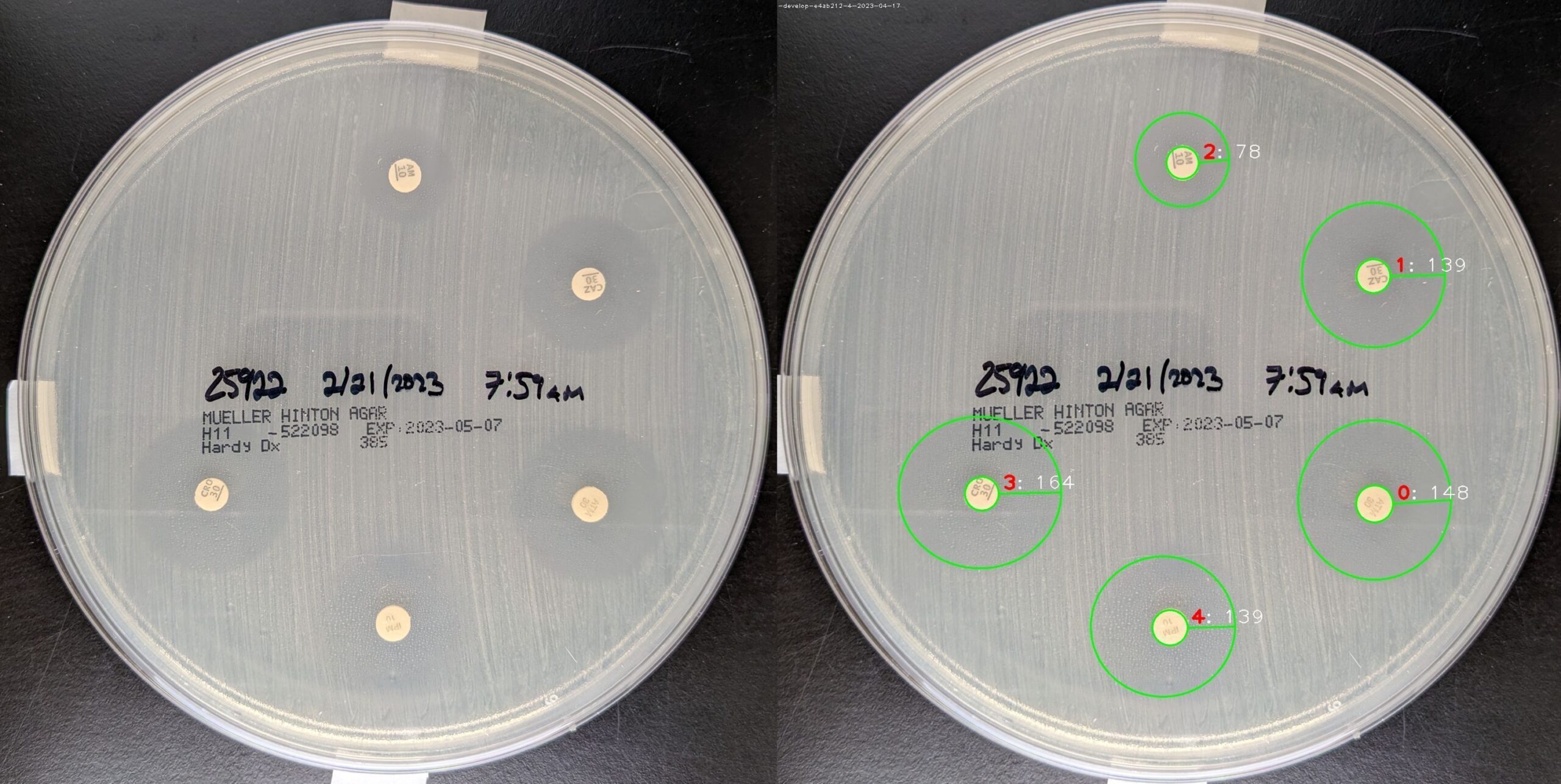
Now, instead of having to use a ruler and the naked eye, you can rely on AI and automation. You can simply take a photo with your smartphone and upload it and all the inhibition zones will be instantly and automatically measured.
This highly accurate design eliminates the issue of human error and saves labs time, money, and energy.
It’s definitely worth checking out.
You can test the Oculyze Disk Diffusion Analyzer for free here. Please note that the recognition provided here is solely for demonstration purposes and may not accurately represent the performance of our product.
If, alternatively, you’re interested in counting the CFUs, check out our Automated Colony Counter.
With our Image Analysis Platform, you can save hundreds of hours of work that would have gone into manual labor. Image analysis automation provides you with accurate results and secure data management in less time, so you can save your resources for something that brings more value to your business.
Let artificial intelligence do the hard work for you. Start saving time and costs now! Want to know more? Contact us and we’ll be happy to help!


